Categories
Normal Variants

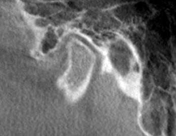
Normal Condyle

Normal condyle should be in the physiological center. The joint space should be 3-5 mm thick. The cortical plate should be smooth and not pitted.

Categories
Uncategorized

3D 4th Dimension Views

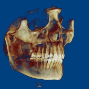
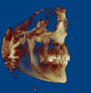
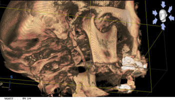
A 3D radiograph with a 4th dimension view of the skull gives a distinct advantage in diagnosis of TMJ, sleep apnea and orthodontic problems.

Please click an image below to see an AVI movie showing the effect of 4th dimension.

Categories
Sleep Apnea

Narrow Airway

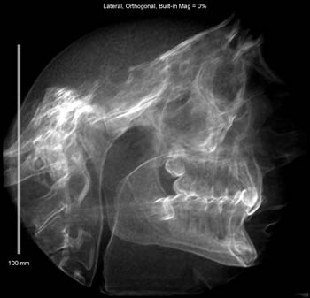
If the mandible is displaced posterior, the tongue can also be displaced posteriorly. The combination of displaced jaw function and posteriorly positioned tongue may be the cause of narrowed airway.

Categories
Global Impression

Overall Appearance

When looking at the radiograph, your first mind image is what is the over all apperance of the radiographic image.
What is the symetry?
What is Density?
What is the overall cortical appearance?

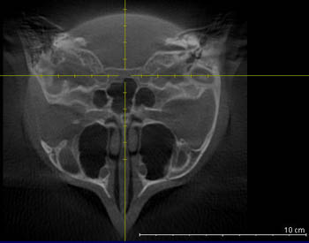
Axial View of skull at the condyle level.

Please email me if any question.

Categories
Local Impression

Local Appearance

Where is the pathology? The condylar problem is usually located at the head of the mandible.
The condylar surface can be flattened or show signs of osteoarthritis.

Please contact me if any question.

Categories
TM Joint

Symptom of Osteoarthritis

Osteoarthritic condyle is a common problem with TM Joint disease.